Home
Uncategories
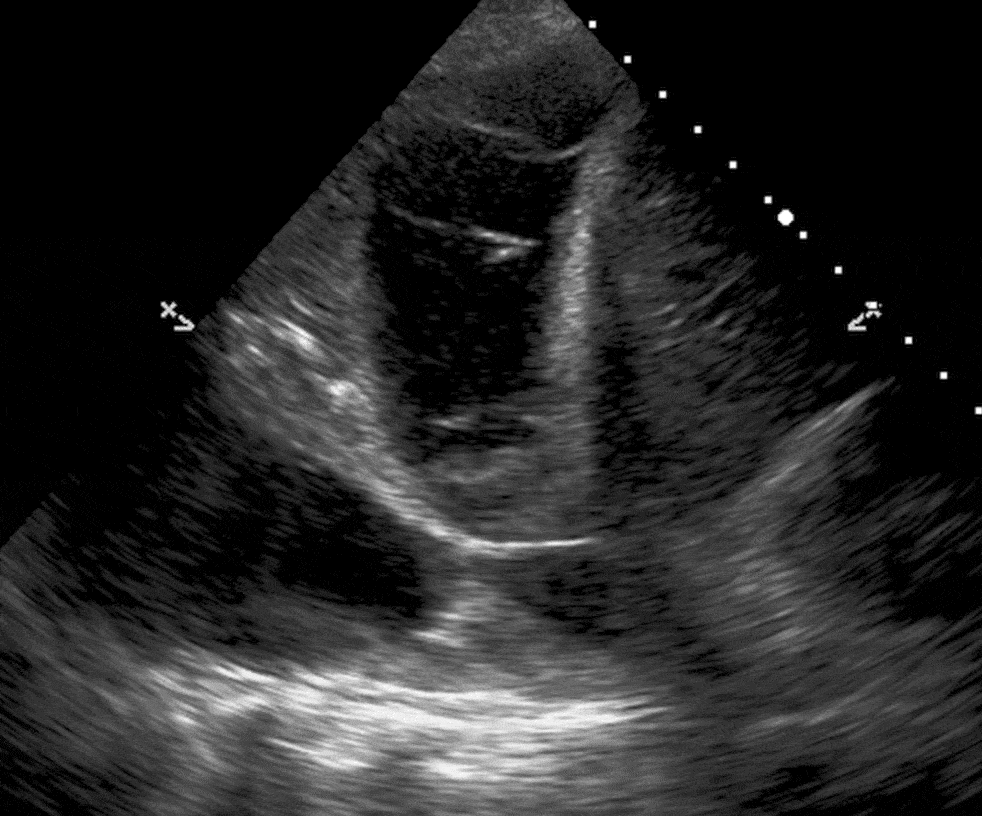
Loculated Pleural Effusion : 2 Lung Ultrasound Pre-Reading for FCUS course - Intensive ... / Learn more about the symptoms of this lung condition and your treatment.
Loculated Pleural Effusion : 2 Lung Ultrasound Pre-Reading for FCUS course - Intensive ... / Learn more about the symptoms of this lung condition and your treatment.
Loculated Pleural Effusion : 2 Lung Ultrasound Pre-Reading for FCUS course - Intensive ... / Learn more about the symptoms of this lung condition and your treatment.. Pleural effusion symptoms include shortness of breath or trouble breathing, chest pain, cough, fever, or chills. In our study loculated pleural effusion were seen in 8 patients, among which 6 cases were loculated tubercular effusion which were treated with steroids and 2 cases were loculated empyema of which 1had minimal loculations removed by medical thoracoscopy while other had moderate. Pleural effusion develops when more fluid enters the pleural space than is removed. The pleura are thin membranes that line the lungs and the inside of the chest cavity and act to lubricate and facilitate breathing. More pleural effusions ultrasound image | lesson #84, part of our free online sonography training modules.
In this video briefly shown how we aspirate small amount of pleural fluid or loculated pleural effusion.for more videos please subscribe the channel.if you. When you have a pleural effusion, fluid builds up in the space between the layers of your pleura. Often it happens in the context of a pneumonia, injury, or chest surgery. Case contributed by dr prashant mudgal. Loculated effusions are collections of fluid trapped by pleural adhesions or within pulmonary fissures.
Lung Ultrasound Made Easy: Step-By-Step Guide - POCUS 101 from pocus101.b-cdn.net An exudative pleural effusion occurs when there is increased permeability of the pleural surface and/or capillaries, usually as a result of inflammation. Pleural fluid ldh > two thirds of upper limit for serum ldh. Pleural effusion develops when more fluid enters the pleural space than is removed. It was successful in breaking the locules. Ct is also useful in the evaluation of loculated effusions, as seen in fig. A role in selected clinical circumstances. Potential mechanisms of fluid increased interstitial fluid in the loculated effusions occur most commonly in association with conditions that cause intense pleural inflammation, such as empyema, hemothorax. Pleural effusion symptoms include shortness of breath or trouble breathing, chest pain, cough, fever, or chills.
Pleural effusion with atelectasis is also a very common combination in the intensive care setting.
Pleural effusions unlikely associated with ra as transudative, and without monocyte predominance or low glucose. Often it happens in the context of a pneumonia, injury, or chest surgery. Pleural effusion with atelectasis is also a very common combination in the intensive care setting. It was successful in breaking the locules. It is one of the various kinds of pleural effusion. Pleural effusions can loculate as a result of adhesions. Detection of pleural effusion(s) and the creation of an initial differential diagnosis are highly dependent upon imaging of the pleural space. Pleural effusion is the accumulation of fluid in the pleural space resulting from disruption of the homeostatic forces responsible for the movement of pleural fluid. Treatment depends on the cause. Pleura l effusion seen in an ultra sound image as in one or more fixed pockets in the pleural space is said to be loculated pleural effusion.in. Pleural fluid/serum protein ratio >0.5. Case contributed by dr prashant mudgal. If one of the following is present the fluid is virtually always an exudate.
Pleural fluid/serum protein ratio >0.5. Obliteration of left costophrenic angle with a wide pleural based dome shaped opacity projecting into the lung noted tracking along the cp angle and lateral chest wall suggestive of loculated pleural. Us scan they can be identified clearly and it is very complicated.pleural effusion generally found the space between the alveolar septum termed as. Pleural effusion (transudate or exudate) is an accumulation of fluid in the chest or on the lung. Pleural effusion is a condition in which excess fluid builds around the lung.
Loculated transudative pleural effusion masquerading as ... from cdn.amegroups.cn Diffuse nodules and opacification in right lung with compressive atelectasis. A pleural effusion is accumulation of excessive fluid in the pleural space, the potential space that surrounds each lung. Pleural effusion, also called water on the lung, is an excessive buildup of fluid between your lungs and chest cavity. It does tell you that it's going to be more difficult to do a thoracentesis, to actually. Transudative pleural effusion, where the excess pleural fluid is low in protein is caused by fluid leaking into the pleural space. Pleural effusions unlikely associated with ra as transudative, and without monocyte predominance or low glucose. When a pleural effusion is loculated, the standard treatment methods of intercostal tube drainage and pleurodesis may not be helpful. Pleural empyema is a collection of pus in the pleural cavity caused by microorganisms, usually bacteria.
If none is present the fluid is virtually always a transudate.
Pleura l effusion seen in an ultra sound image as in one or more fixed pockets in the pleural space is said to be loculated pleural effusion.in. Pleural effusion is the accumulation of fluid in the pleural space resulting from disruption of the homeostatic forces responsible for the movement of pleural fluid. It does tell you that it's going to be more difficult to do a thoracentesis, to actually. Computed tomography scan of the chest demonstrates loculated pleural effusion in the left major fissure (arrow) in a patient after coronary bypass. When a pleural effusion is loculated, the standard treatment methods of intercostal tube drainage and pleurodesis may not be helpful. Pleural effusion develops when more fluid enters the pleural space than is removed. Pleural fluid/serum ldh ratio >0.6. Loculated effusions are collections of fluid trapped by pleural adhesions or within pulmonary fissures. A role in selected clinical circumstances. Learn more about the symptoms of this lung condition and your treatment. Pleural fluid/serum protein ratio >0.5. Often it happens in the context of a pneumonia, injury, or chest surgery. It is one of the various kinds of pleural effusion.
An exudative pleural effusion occurs when there is increased permeability of the pleural surface and/or capillaries, usually as a result of inflammation. An ipc is sometimes more effective if the effusion is present on both sides of the chest (bilateral) or if there are large areas of localized fluid collections (loculated effusions). Learn more about the symptoms of this lung condition and your treatment. Pleural effusions can loculate as a result of adhesions. Treatment depends on the cause.
Next from www.meddean.luc.edu Treatment depends on the cause. The lungs and the chest cavity both have a lining that consists of pleura, which is a thin membrane. The pleura are thin membranes that line the lungs and the inside of the chest cavity and act to lubricate and facilitate breathing. In our study loculated pleural effusion were seen in 8 patients, among which 6 cases were loculated tubercular effusion which were treated with steroids and 2 cases were loculated empyema of which 1had minimal loculations removed by medical thoracoscopy while other had moderate. The pleura is a thin membrane that lines the surface of your lungs and the inside of your chest wall. Diffuse nodules and opacification in right lung with compressive atelectasis. Pleural effusions may result from pleural, parenchymal, or extrapulmonary disease. Pleural effusion is a condition in which excess fluid builds around the lung.
Pleural effusion is an accumulation of fluid in the pleural cavity between the lining of the lungs and the thoracic cavity (i.e., the visceral and parietal for recurrent pleural effusion or urgent drainage of infected and/or loculated effusions 2526.
Pleural effusions unlikely associated with ra as transudative, and without monocyte predominance or low glucose. Pleural fluid/serum protein ratio >0.5. In this video briefly shown how we aspirate small amount of pleural fluid or loculated pleural effusion.for more videos please subscribe the channel.if you. Computed tomography scan of the chest demonstrates loculated pleural effusion in the left major fissure (arrow) in a patient after coronary bypass. A malignant pleural effusion may be large and diffuse or small and involve just a small portion of the pleural cavity. Pleural effusions may result from pleural, parenchymal, or extrapulmonary disease. Diffuse nodules and opacification in right lung with compressive atelectasis. Pleural effusions can loculate as a result of adhesions. Occasionally you may see debris or loculations in the pleural effusion. Pleural fluid ldh > two thirds of upper limit for serum ldh. Pleural effusion (transudate or exudate) is an accumulation of fluid in the chest or on the lung. Case contributed by dr prashant mudgal. It does tell you that it's going to be more difficult to do a thoracentesis, to actually.
This is a short description in the author block about the author. You edit it by entering text in the "Biographical Info" field in the user admin panel.


0 Tanggapan:
Post a Comment